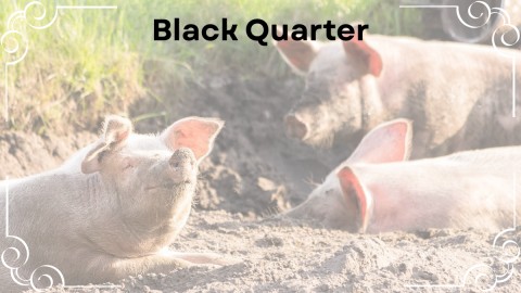
This is an acute infectious but not contagious disease of cattle, goat and sheep. The disease is characterized by development of focal gangrenous and emphysematous myositis. This gives rise to crepitation and sero harmorrhagic swelling in the heavy muscles like gluteal muscles. The disease produces severe toxaemia with a very rapid course and high mortality. Black quarter is a predominant disease of cattle but traumatic black quarter may be seen in other animals.
Distribution
The disease is wide spread in most of the tropical countries of the world. In some regions, the disease may remain confined due to contamination of the soil surfaces where as in other parts the disease may be seasonal in distribution. The disease spreads rapidly following heavy rainfall. The disease is most commonly seen in areas where well developed cattle and sheep rearing programmed are in existence.
The disease was first recognised by Billinger in 1975. The disease is wide spread in USA. Breed (1937) and Vowter (1942) remarked that in USA, the disease was caused by Clostridium septicum and Cl. chauvoei.
In India, The disease is sporadic in nature. Infection rate to the extent of 85% has been recorded from Madras, Bombay, Mysure and Hydrabad and only 15% from rest parts of the country. The distance appears in almost all the states of the country during rainy season as a seasonal entity excepting Madras, where the disease is evenly distributed all round the year. The disease is prevalent in all the states of India as a sporadic problem.
Aetiology
Black quarter is caused by Clostridium chauvoei, a gram positive rod shaped, spore forming toxin producing anaerobe. They are 0.6 micron in diameter and 3 to 8 micron in length.
The spores are very much resistant to altered environmental stress. The spores are very much unaffected to hot, dessication and disinfectants. Spores can withstand 120° C temperature for 10 minutes. The pores can persist in the soil for number of years. The bacteria may remain viable for more than eight years in dried up muscle tissues. The organisms can be destroyed by 3% formalin in 15 minutes and by 2% bichloride of mercury in 10 minutes.
False black quarter may be caused by Cl. Septicum and Cl. novyi. These organisms may have role in the pathogenesis of the disease. But, the importance of Cl. septicum as a cause of the disease is questionable. In smers made from the surface of liver of infected guinea pig Cl. chauvoei remains as single bacilli or short chain while Cl. septicum forms chain. Cl. chauvoei has got one biotype and it is serologically homogenus where as Cl. septicum has got four major anigenic groups. They produce potent exotoxin, haemolyse erythrocytes and remain stable when exposed to air. In a study in India from cased of black quarter in cattle and buffalo from all over the countries 90.5% were typed as Cl. chauvoei, 5.6% as Cl. septicum and 2.2% as mixtures of Cl. chauvoei and Cl. septicum, Cl. chauvoei is considered as the true cause of black quarter and other anrobes isolated from the disease are thought to be post mortem invaders.
Susceptible Hosts
Cattle is the most susceptible host but the infection may spread to other animal due to traumatization of the muscles. The disease may occur in sheep, buffalo and goat. The disease has been recorded in deer. A horse has been found to suffer. In a very rare occasion pig has been found to be affected.
Cattle of all breeds are susceptible but he incidence is more in cattle having 4 to 24 months of age group and good body condition. Of the laboratory animals, guinea pigs and rabbits are most susceptible.
Mode of Transmission
The disease spreads from contaminated soils. The contamination of soil is due to infected carcases which cause pollution of the land. The organisms gain entry through ingestion of infected feeds or possibly through contamination of wounds. The normal healthy animals may harbour bacteria in the spleen, liver and alimentary tract and thus the animals may excrete the organisms through their faeces. In sheep, the bacteria is introduced through shearing, docking, lambing and castration. In rare occurrence, the disease has been found to flare up in sheep. Wounds due to fighting, naval infection, vulva and vaginal infection at birth may cause local lesions. Nilkanthan and Dhanda (1959) could experimentally transmit the disease in guinea pig.
Pathogenesis
Ingested organisms are carried from the intestine via circulation to the skeletal muscles. The spores from the alimentary tract penetrate tissues from the places of breach of alimentary mucosa due to trauma. Some of the spores in the muscles are destroyed by phagocytosis and others remain latent for at least several weeks. Very often, well-formed heavy muscles like muscles of the gluteal region, loin, shoulders are affected. The infection may affect the muscles and intermuscular tissues and blood capillaries. Gases used to accumulate within the muscle fibres due to fermentation. Haemorrhages from ruptured capillaries may occur. The muscles of the tongue, jaw, heart may be affected at a later stage due to dissemination of infection. The infection can also be spread through peritoneum and pleura. Exotoxin is produced from the organisms which causes systemic reaction characterized by toxaemia and local reaction characterized by necrotizing myositis. Severe toxaemia may lead to death. Following death there is rapid tissue decomposition and spores are formed in the dehydrated tissues. Recovered animals are immune. Smith (1980) opined that Cl. chauvoei is not a potent exotoxin former. But, highly virulent strains have been regarded to produce exotoxin which could produce oedema in guinea pig and oedema along with pulmonary congestion in mice on subcutaneous inoculation.
Clinical Findings
The incubation period is usually 2 to 5 days. Under the influence of toxic products elaborated during the growth of the organisms, the muscles degenerate and gases are evolved. The toxic products are absorbed by the body fluids causing systemic disturbances and a decrease in the animals vitality.
Cattle
In cattle, the first symptom is a rise in body temperature which may be as high as 106° C or 108° C, but sometimes, there is hardly any sign of fever. The appetite is lost and rumination is suspended and there is stiffness or lameness in one of the limb. This being the usual early symptom. Very soon characteristic swelling develops in one of the thick layers of muscles. Most commonly the lesions are located in thigh, buttocks, shoulder, neck and lumber region and more rarely in the inter-mandibular space or in the tongue. Swelling are hot and painful in the early stage and become cold and painless latter. The muscle tissues are swollen, dark in colour and turn dry. The structures get disrupted due to gas pocket giving rise to a spongy texture. On pressure swellings emit crackling or crepitation sound due to emphysema. Skin over the swelling is discoloured and dry. There is laboured breathing and accelerated pulse rate (100-120/minutes). Occasionally, there is distinct abdominal pain. Finally, the temperature drops and the patient dies within 12 to 48 hours after manifestation of clinical signs.
Sheep
In sheep, there is extensive haemorrhagic oedema in the subcutaneous tissues following a wound. Lameness is not a constant feature. There is complete loss of appetite, high rise of temperature, profound depression and death. Lameness may be present in some sheep for which it may not be able to stand and walk. The animal used to die within 24 to 48 hours post infection.
Horse
In horse, oedema of the pectoral muscles, stiffness of gait and ataxia have been observed.
Lesions
Lesions are limited to affected muscles. Muscles of shoulder, thigh and neck are usually affected. Lesions may also be observed in the tongue, diaphragm and myocardium. Large crepitating swelling are the most characteristic necropsy findings. Affected muscles are infiltrated with yellowish exudate. Gas bubbles accumulate between the muscle fibres. Due to haemorrhage affected tissues turn black. A rancid odour emanates from the muscles. Blood stain discharge may ooze from the nostrils. There is accumulation of fluid in the pericardium. Heart muscles remain blackish red and there is parenchymatous degeneration. The conjunctival mucous membrane become congested. The liver, kidneys, lungs and spleen show yellowish foci and haemorrhage. The entire body assumes a bloated appearance. Putrefaction and bloating occur very quickly. In some cases, bloodish frothy discharge from natural orifices and prolapse of the rectum may be evident.
On microscopical examination, the affected muscles show coagulative necrosis, infiltration of leukocytes and haemolysis of erythrocytes. Some muscle bundles are separated by gas bubbles. Muscles may reveal necrosis and fibro-haemorrhagic exudation. Gram +ve rods are noted throughout the lesions.
Diagnosis
In the field outbreak, a tentetive diagnosis is made from the history, clinical observation and postmortem findings.
In the laboratory, the disease may be diagnosed by the following methods.
Microscopical Examination of Smear
By examination of smear made from the affected tissues or fluids of the swellings, gram positive rods with subterminal spores will be seen. Sometime, Cl. chauvoei and Cl. septicum may remain as mixed infection. Therefore, attempt should be made for specific isolation and identification to have definite diagnosis of Cl. chauvoei.
Cultural Test
Anaerobic media provides growth within 23 hours. Materials to be used for cultural examinations are heart blood, peritoneal fluids affected muscles either from the carcase of natural host or from the inoculated laboratory animals.
Biological Test
Muscle pieces from the lesions of the dead animal are cut up into small pieces, triturated with sterile saline and filtered. The filtrates containing spores are heated to 60° C for 30 minutes. One ml of these are injected into the gluteal muscles of guinea pig. In positive cases where the materials contain spores of Cl. chauvoei, the animals die within 48 hours. Liver impression smears reveal numerous organisms which are gram +ve having subterminal spores.
Fluorescent Antibody Technique
This is useful for correct diagnosis and also to identify mixed infections. Fluorescent labelled specific antibodies are added to the infected tissues, exudates or cultures those are fixed on slides. Then the slides are exposed to fluorescent antibody for 30 minutes. Then the slides are washed in phosphate buffer for 10 minutes and mounted in 90% buffered glycerol. The slides are then examined under a microscope adopted for fluorescent microscopy. Specific organism is indicated by marked fluorescent.
Differential Diagnosis
Black quarter must be differentiated from anthrax, malignant oedema and bacillary haemoglobinuria.
Anthrax and bacillary haemoglobinuria may be enzootic in some places and may occur at any season of the year. Bloating and bloody discharges may be seen in animals those die of these diseases in warm weather. But, subcutaneous crepitation and local swellings are absent in animals which die only a few hours back. Black quarter must not be confused with wet clover poisoning, a non-febrile, non-transmittable disease of cattle, which is characterized by excessive fluctuating haemolytic swelling that may appear on any superficial part of the body especially in the gluteal, costal or shoulder regions. These swellings contain large amount of blood. They pit on pressure and crepitation is absent.
Treatment
Satisfactory response has been reported from the use of penicillin, aureomyin and oxyetracycline. The antibiotic may be injected into the affected muscles. Penicillin is extensively used and considered as drug of choice. Penicillin @ 2000 to 4000 units per pound body weight per day may be used. Crystalline penicillin may be given through intravenous route followed by procaine penicillin through intramuscular route. Newer antibiotic e.g. Cephaloridine may be tried.
Control
Since the disease is associated with infection from the soil, the cultivation in that soil may be avoided.
- The young animals should be kept out of such area.
- The dead body should be burnt or buried.
- The dead body should not be allowed to skin.
- The calf and sheep should not be allowed to graze in endemic pasture.
- All the animals of the endemic zones should be vaccinated with suitable vaccine.
- Hogerth-Scott (1980) recommended the use of polyvalent vaccine and anthelmintic combination to control the black quarter in a flock of sheep.

